Friday, July 15, 2016

FEVER IN A CHILD

Fever in a Child

is the most common chief complaint presenting to an emergency department and accounts for 30% of outpatient visits each year. Early studies suggested that infants younger than 3 months were at high risk of a serious bacterial illness (SBI), which included sepsis, pyelonephritis, pneumonia, and meningitis. Current practice guidelines vary in their cut-offs for evaluation and treatment strategies. Neonates are clearly at the highest risk, while infants in their second and third months of life gradually transition to the lower risk profile of older infants and children. The incidence of bacteremia falls from around 10% among febrile neonates to approximately 0.2% in immunized infants and children older than 4 months; meningitis risk decreases from about 1% in the first month of life to < 0.1% later in infancy; the risk for pyelonephritis remains relatively constant among young girls with fever, and gradually decreases among boys over the first year of life. The individual practitioner must weigh these risks against the invasiveness of their ED evaluation and make shared decisions with the family on the best approach.

CLINICAL FEATURES:

In the neonate or infant < 2 to 3 months of age, the threshold for concerning fever is 38°C (100.4°F); in infants and children 3 to 36 months old, the threshold is 39°C (102.2°F). In general, higher temperatures are associated with a higher incidence of serious bacterial illness.
Young infants are especially problematic in assessing severity of illness. Immature development and immature immunity make reliable examination findings difficult. Persistent crying, inability to console, poor feeding, or temperature instability may be the only findings suggestive of an SBI.

Sunday, May 15, 2016

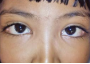
Epiblepharon & Euryblepharon

Epiblepharon


 Extra fold of skin in the lower lid with inturning of eyelashes

 Nasal 1/3rd is most commonly affected

 Treatment: Plastic repair if necessary to prevent recurrent infection.

Euryblepharon

Rare, congenital, bilateral, not so serious condition


  •  Palpebral apertures are larger than normal 


              and may be with epicanthus


  • Excessive watering may be a problem due to more exposure


 Treatment:

 No treatment for most of the cases; lateral tarsorrhaphy for symptomatic cases.

Bilateral eyelids drooping Disease

Blepharophimosis Syndrome (Bilateral eyelid drooping disease)

 Autosomal dominant Condition
(Can be transmitted from both mother or father, usually express in every generation)


Features:

Syndrome consists of bilateral:

– Narrowing of vertical and horizontal palperbral apertures

– Telecanthus

– Inverse epicanthus folds

– Lateral ectropion and moderate to severe ptosis

 May be asymmetrical and without epicanthic folds


 Treatment: 

Plastic reconstruction of lids, along with bilateral brow suspension for ptosis.

Distichiasis

Distichiasis

 Hereditary and congenital condition

 Extra posterior row of cilia, occasionally present in all four lids

 Partial

Complete 


 They occupy the position of meibomian gland orifices

 Eyelashes may irritate cause to corneal epithelial defects

 May be also seen in Stevens Johnson’s syndrome—acquired distichiasis


 Treatment:

By cryotherapy or excision with grafting.

Congenital Lid Conditions: Ophthalmology Spot Diagnosis Series

Coloboma of the Eyelid

 A notch or defect of the lid margin

 Unilateral 

 or bilateral ; upper or lower

 Upper lid coloboma: At the junction of middle and inner thirds

 May be associated with Goldenhar syndrome

 Lower lid coloboma: At middle and outer thirds junction


 Associated with Treacher Collins syndrome

 May be acquired in traumatic cases


 Treatment: 

Urgent plastic repair at a very early age to prevent exposure keratitis and corneal ulcer.

Tuesday, May 10, 2016

Vertigo: what are the causes!

Vertigo

Most important causes of vertigo

DisorderNotes
Viral labyrinthitisRecent viral infection
Sudden onset
Nausea and vomiting
Hearing may be affected
Vestibular neuritisRecent viral infection
Recurrent vertigo attacks lasting hours or days
No hearing loss
Benign paroxysmal positional vertigoGradual onset
Triggered by change in head position
Each episode lasts 10-20 seconds
Meniere's diseaseAssociated with hearing loss, tinnitus and sensation of fullness or pressure in one or both ears
Vertebrobasilar ischaemiaElderly patient
Dizziness on extension of neck
Acoustic neuromaHearing loss, vertigo, tinnitus
Absent corneal reflex is important sign
Associated with neurofibromatosis type 2

Sunday, January 31, 2016

Abdominal Aortic Aneurysm

Abdominal Aortic Aneurysm


Basics
Description
  • Focal dilation of the aortic wall with an increase in diameter by at least 50% (>3 cm)
  • 95% are infrarenal.
  • Gradual expansion or rupture causes symptoms.
  • Rupture can occur into the intraperitoneal or retroperitoneal spaces.
  • Intraperitoneal rupture is usually immediately fatal.
  • Average growth rate of 0.2 to 0.5 cm per year
  • 5-year risk of rupture:
    • Aneurysms <4.0 cm: 2%
    • Aneurysms 4.0-5.0 cm: 5%
    • Aneurysms 5.0-6.0 cm: 25%
    • Aneurysms 6.0-7.0 cm: 35%
  • 40-50% die before they reach the hospital.
  • 50% of patients who reach the hospital alive survive.
  • 5-year survival after repair is 67%.
Geriatric Considerations
  • Risk increases with advanced age.
  • Present in 4-8% of all patients older than 65 years
  • Peak incidence:
    • Men: 5.9% at the age of 80 years
    • Women: 4.5% at the age of 90 years
Etiology
  • Risk factors:
    • Male gender
    • Age >65 years old
    • Family history
    • Cigarette smoking
    • Atherosclerosis
    • Hypertension
    • Diabetes mellitus
    • Connective tissue disorders:
      • Ehlers-Danlos syndrome
      • Marfan syndrome
  • Uncommon causes:
    • Blunt abdominal trauma
    • Infections of the aorta
Related Posts Plugin for WordPress, Blogger...