Showing posts with label Guidelines. Show all posts

Monday, March 23, 2020

How chloroquine will act as a prophylactic drug and is acting as a therapeutic agent against COVID-19 infection

*How chloroquine will act as a prophylactic drug and is acting as a therapeutic agent against COVID-19 infection.*

Chloroquine acts on host target respiratory cells by:
Chloroquine increases the endosomal pH required for the virus- host target cell fusion. Increase in the pH disrupts the normal viral function.
In SARS-coronavirus, which is a sister to COVID-19, chloroquine was found to interfere with the glycosylation of cellular receptors of the virus. This interference eventually resulted in no association between the host target cell and the virus.
Chloroquine acts as an ionophoric agent for Zinc ions and thus increases the in flux of zinc ions into the cytoplasm of host target cells regardless whether the host target cells are infected or not.

All the above-mentionedgas mechanism is on the host cells and COVID-19 can not mutate and cause resistance to these 3 mechanisms. First 2 mechanisms inhibit the virus-target host cell union. Chloroquine results in disablement of ACE2 (Angiotensin converting enzyme 2) terminal glycosylation which leads to the morphological change; ACE-2 is a surface receptor found on target host cell. This results in the disruption in the association between the COVID-19 and target host cell as COVID-19 requires ACE-2 receptor to attach to a cell.     
Because the action is on the target host cell, Chloroquine won’t develop resistance therapeutically. If a person uses Chloroquine as a prophylactic agent (500mg once in a week for adults and 8.3 mg per kg once in a week for children) against COVID-19 then, it will act pre-infection and post-infection. If a person does not get exposed to the COVID-19 infection after taking Chloroquine (say for 3 weeks) and then some viruses enters and try to infect target host cells, Chloroquine mechanism “a” and mechanism “b” will prevent the union of virus-target host cell. If some of the viruses enters the target host cell, there Zinc ions are waiting to adhere to the RNA dependent RNA polymerase enzyme of the virus and stops COVID-19 polymerization intracellularly. If COVID-19 mutates inside the cell several times, even then the Zinc ions will actively inhibit the viral multiplication inside the host respiratory cells, irrespective of the viral strain. Even if COVID-19 virus manages to escape from Zinc ions trap and releases from the host target cell cytoplasm into the interstitial matrix, intercellular space, and tried to re-infect some of the healthy target host cells, Chloroquine will prevent the re-union of viral genome with target host cells via mechanism ‘a’ and mechanism ‘b’ and the infection will halt in the preliminary stages itself and complications like COVID-19 pneumonia will not develop. Chloroquine molecules will not lose its effectivity in an individual pre and post infection.
Zinc is present in ample amount in the human body. In normal conditions, the Zinc is not present in free state in the cell. The increased level of Zinc ions is not toxic to the cells and cells excrete the extra Zinc ions into the extracellular space. Zinc is commonly present in red meat, legumes, nuts, milk, cheese, eggs, whole grains etc. Garlic increases the absorption and bioavailability of Zinc inside the body. So, some persons who aren’t taking Zinc rich foods in ample amount should eat zinc supplements or garlic daily. 

Safety of Chloroquine is well tested as it is given for
Malaria prophylaxis
Autoimmune diseases like rheumatoid arthritis
Lupus erythematosus
Now a days it is indicated into the SARS infection as it is a broad antiviral agent.
Through it ionophoric action for its zinc intracellular influx, chloroquine is also used as anti-cancerous drug.

The long-term use of chloroquine (4 years together) may cause its accumulation in the eye. There are some concerns of its use in glucose-6-phosphate dehydrogenase enzyme deficient children but the recommended prophylactic and therapeutic doses, Chloroquine is safe to be used in these children. Some persons may complain of acidity and nausea, but it can be resolved if Chloroquine is taken post meal.


Prophylactic dose in malaria is 500 mg once a week in adults and 8.3 mg per kg once a week in children.
In these doses it will be effective against COVID-19 prophylaxis. The therapeutic doses against COVID-19 as used in China, America and India are Chloroquine 500mg BD for 5 days with other anti-viral drugs like Oseltamivir, Lopinavir, Ritonavir etc. and in complicated COVID-19 pneumonia cases, Chloroquine may be used for 10 or more days in the same amount.

Now comes the actual catch of its technicality to be used prophylactically against COVID-19 action. In India as it is a very cheap, safe and easily available drug and can be prescribed against the Malaria as a prophylactic drug as the summer season approaches. It will surely work prophylactically against the COVID-19 outbreak and we can use this opportunity to prescribe it in larger amount as Malaria is endemic in India. It will act on Malaria and COVID-19 infection prophylactically without it being included in the guidelines.
Trump’s excitement for chloroquine in his announcement can be related with the fact that American researchers are currently working on the Chloroquine drug. Many companies have donated chloroquine to the US for research purpose.
Hydroxychloroquine is less toxic but original Chinese work is based on chloroquine phosphate. Hydroxychloroquine can be used with equal efficacy.
If one starts prophylactically with Chloroquine, then one must stick with chloroquine and must not switch to hydroxychloroquine and vice-versa. This switch may result in increase QT interval.

*REFERENCES*
Zhu N, Zhang D, Wang W et al. A novel coronavirus from patients with pneumonia in China, 2019. N Engl J Med 2020
Gao, J., Tian, Z. and Yang, X., 2020. Breakthrough: Chloroquine phosphate has shown apparent efficacy in treatment of COVID-19 associated pneumonia in clinical studies. BioScience Trends.
Vincent, M., Bergeron, E., Benjannet, S., Erickson, B., Rollin, P., Ksiazek, T., Seidah, N. and Nichol, S., 2005. Virology Journal, 2(1), p.69.
https://youtu.be/U7F1cnWup9M


SARS-CoV-2 RNA more readily detected in induced sputum than in throat swabs of convalescent COVID-19 patients - The Lancet Infectious Diseases
https://www.thelancet.com/journals/laninf/article/PIIS1473-3099(20)30174-2/fulltext

Saturday, March 21, 2020

‏LABORATORY MARKERS IN COVID-19

‏LABORATORY MARKERS IN COVID-19 PATIENTS :microscope::syringe:
‏MOST FREQUENT
‏Decrease lymphocyte count
‏Decrease albumin
‏Decrease hemoglobin levels
‏Increase C-reactive protein (CRP)
‏Increase Erythrocyte Sedimentation Rate (ESR)
‏Increase Lactate Dehydrogenase (LDH)
‏Increase D-dimer

- Deaths from Covid-19 are due to (1) cytokine storm syndrome and (2) fulminant myocarditis.

- During a cytokine storm, an excessive immune response ravages healthy lung tissue, leading to acute respiratory distress and multi-organ failure.

- All Covid-19 patients sick enough for hospitalization should be given a serum ferritin blood test.

- Elevated serum ferritin values are a good first screening tool for the possibility of a cytokine storm syndrome in sick patients with high fevers.

- Treating Covid-19 infected patients with cytokine storm syndrome with IL-6 blockade (tocilizumab) has recently been reported in China with successful outcomes.

- Fulminant myocarditis :heart: has also been reported in patients with Covid-19

- Fulminant myocarditis is primarily caused by infection with viruses. It arises quickly, progresses rapidly, and may lead to severe heart failure or circulatory failure presenting as rapid-onset hypotension and cardiogenic shock, with mortality rates as high as 50%–70%.

- Physicians should pay attention not only to the symptoms of respiratory dysfunction but also the symptoms of cardiac injury.


_\\\\______
‏Source: Lippi G, Plebiani M. Laboratory
abnormalities in patient with COVID-19 infection. Clin Chem Lab Med. 2020 Mar 3. doi: 10.1515/cclm-2020-0198 Covid-19 🦠 Update:*

Friday, March 20, 2020

Current criteria to swab for COVID-19

Current criteria to swab for COVID-19 in the UK:

Clinical : Pneumonia clinical or radiological, ARDS, Flu like illness (fever, cough whether productive or non productive, sore throat, runny nose, myalgia. Less people present initially with diarrhea 10% and they had the worst prognosis later from the Wuhan experience)
Or Epidemiological : last 14 days in tier 1 or tier 2 countries. Or contact with confirmed case of COVID-19.

-Incubation period 4 - 14 days. People are infective mostly in the first 5 days when the symptoms are milder. Deterioration happens after day 10 of the symptoms (when the body enter into the adaptive immunity stage)

-PCR for COVID19 sensitive for 70% only, may be initially negative, a repeat PCR in high suspected cases recommended in 48 -72 hours.

-CT Thorax is sensitive for 97% of cases and could help in diagnosis of patient with initial negative PCR (positive CT Thorax for COVID-19 is patchy ground-glass appearance in the lung peripheries, it does not cause cavitation or lymphadenopathy)

- Old age >70, High SOFA score, and d-dimers >1000 are risk factors for poor prognosis.
Pediatrics guidelines if needed please contact me freely and i will provide you details with evidence.

-Lymphopenia with normal WBC count is common in 80%, mild thrombocytopenia , high CRP and ferritin tract with disease severity.

-High tropronin found in half of the dead patients (perhaps as IHD was a common risk factor, likely it is troponin leakage form sepsis /strain? some reported cases for severe myocarditis). Considered as a marker for poor prognosis.

-Give empirical antibiotics in the golden hour as per the normal sepsis protocol . Blood cultures./Procalcitonin. Review ABX in 48 hours.
-NO IV Fluids, unless there is an evidence of hypo-perfusion.
-No role of steroids as it delays viral clearance, unless the patient has obstructive airway disease or Septic shock not fluid or vasopressor responsive

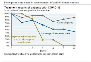
-Controversial role of Hydroxychloroquine: it interfers with cellular ACE receptors in the lung, hence has some anti-viral activities.

-Some Antiviral therapies like Remdesivir, combination of Lopinavir/Ritonavir/Ribavirin is of benefit in MERS, no evidence of use it in COVID-19, Clinical trials are taking place currently. If the patient deteriorated, consider them after discussion with the local ID team. No role of using Oseltamavir in COVID-19 unless influenza is considered when you admit the patient.

- Cytokines storm inhibitors in COVID-19: as the disease cause a cytokines storm (elevated inflammatory markers like CRP, ESR, ferritin), selective inhibition of cytokines Tocilizumab showed some benefit in phase 3 RCT in China. The drug was eventually licensed in China for severe COVID-19 infection.

- COVID-19 may cause secondary HLH?

-Severe respiratory failure/?ARDS: ITU for early Intubation and ventilation with low TV and allow for permissive hypercapenia, prone, paralysis, APRV, ECMO.
(HFNO and NIV is not recommended)

-Cardiac arrest in COVID-19:
Identify resuscitation status for any ?COVID-19 hospital admission.
Recognize cardiac arrest by absence signs of life and absent carotid pulse. Do not do listen and feel for breathing.
Wear full PPE (FFP3, Visor, Gloves, plastic apron) then start CPR. The likely cause is a hypoxic arrest. However, the recommendation from resuscitation council UK is to start with CRP. Early ventilation is advised.


Thursday, January 19, 2017

qSOFA: UPDATED 2017

This is the latest Published article so far:

Sepsis: qSOFA More Accurate Than Previous Criteria in ED
Nicola M. Parry, DVM
January 19, 2017

In the emergency department (ED) setting, the quick Sequential Organ Failure Assessment (qSOFA) score is better than previous criteria for predicting in-hospital mortality among patients with suspected infection, a new study suggests.

Yonathan Freund, MD, PhD, from Pitié-Salpêtrière University Hospital, Paris, France, and colleagues published the results of their multicenter prospective cohort study online January 17 in JAMA.

"Among patients presenting to the [ED] with suspected infection, the use of qSOFA resulted in greater prognostic accuracy for in-hospital mortality than did either SIRS [systemic inflammatory response syndrome] or severe sepsis [criteria]," the authors write.

In 2016, a task force of experts was convened to redefine sepsis in the Third International Consensus Definitions for Sepsis and Septic Shock (Sepsis-3). In the previous guideline, the definition of sepsis was based on SIRS criteria, which are nonspecific. In Sepsis-3, the experts redefined sepsis as life-threatening organ dysfunction resulting from a dysregulated host response to infection.

In patients with infection, organ dysfunction is now be identified by an increase in the SOFA score of at least 2 points. The experts recommend that clinicians use qSOFA in settings such as the ED to more rapidly identify patients with sepsis.

Dr Freund and colleagues therefore set out to prospectively validate qSOFA as a mortality predictor in the ED and to compare the accuracy of the new criteria with both the previously used SIRS criteria and the previous definition of severe sepsis (namely, at least two elements of SIRS and a blood lactate level of more than 2 mmol/L (18 mg/dL).

They conducted the study during a 4-week period in 30 EDs in France, Switzerland, Spain, and Belgium, including adult patients with a suspicion of infection who visited the recruiting EDs. Patients were screened and followed until death or hospital discharge.

For each patient, the ED physician recorded the three components of the qSOFA at their worst level during the ED stay (highest respiratory rate, lowest systolic blood pressure, and lowest Glasgow Coma Scale score). The presence of an altered mental status was also recorded independent of Glasgow Coma Scale.

The analysis included 879 patients with a median age of 67 years; 414 (47%) were women. Respiratory tract was the most common site of infection (n = 379; 43%).

"Overall, in-hospital mortality was 8%," the authors note. Patients with a qSOFA score lower than 2 had a 3% mortality rate (95% confidence interval [CI], 2% - 5%), whereas those with a score of 2 or higher had a 24% mortality rate (95% CI, 18% - 30%; absolute difference, 21% [95% CI, 15% - 26%]).

The researchers found qSOFA to be better at predicting in-hospital mortality (area under the receiver operating curve [AUROC], 0.80; 95% CI, 0.74 - 0.85) than the SIRS criteria (AUROC, 0.65; 95% CI, 0.59 - 0.70) and severe sepsis criteria (AUROC, 0.65; 95% CI, 0.59 - 0.70; P < .001 compared with qSOFA).

qSOFA had a sensitivity of 70% (95% CI, 59% - 80%) and specificity of 79% (95% CI, 76% - 82%) for prediction of in-hospital mortality. The test had a positive likelihood ratio of 3.40 (95% CI, 2.80 - 4.17) and a negative predictive value of 97% (95% CI, 95% - 98%).

After adjusting for patient age and site of infection, a qSOFA of 2 or higher was associated with in-hospital mortality with a hazard ratio (HR) of 6.2 (95% CI, 3.8 - 10.3; Harrell C, 0.83). In comparison, using the previous definition of severe sepsis, the HR was 3.5 (95% CI, 2.2 - 5.5).

"One of the strengths of this study is that it prospectively validates the [task force's] findings and highlights how these findings particularly apply to ED patients with even stronger results," the authors conclude.

An accompanying editorial emphasizes the importance of timely identification of patients with possible sepsis.

"[C]linicians must rely on clinical judgment, potentially augmented by clinical criteria validated to identify sepsis among patients with infection," write François Lamontagne, MD, from the University of Sherbrooke, Canada, and David A. Harrison, PhD, and Kathryn M. Rowan, PhD, both from the Intensive Care National Audit & Research Centre, London, United Kingdom.

According to the editorialists, studies have mainly used predictive validity to assess qSOFA. They highlight one particular study of patients with suspected infection that compared use of qSOFA with use of the more complex SOFA or Logistic Organ Dysfunction System tests for patient encounters in hospital settings. In the intensive care unit setting, qSOFA had a worse predictive validity for in-hospital mortality than SOFA or the Logistic Organ Dysfunction System had. However, outside the intensive care unit, qSOFA had a similar or better predictive validity than the other two tests had, despite its relative simplicity.

Although the editorialists acknowledge that qSOFA could therefore be most useful outside the intensive care unit, they stress that the test still needs further evaluation in other settings. For example, in some settings, factors such as how quickly patients with a suspected infection present to the hospital might vary substantially and could affect the test's predictive validity.

Dr Lamontagne and colleagues also suggest that use of qSOFA could be replaced by any highly accurate, rapid diagnostic tests for sepsis that might emerge. However, at least for now, "qSOFA appears a simple, rapid, inexpensive, and valid way to identify — among patients with suspected infection — those at a higher risk of having or developing sepsis," they conclude.

Dr Freund received lecturer fees from Daichi-Sankyo. One coauthor received personal fees from ThermoFisher Brahms, CrossJect, Sanofi, and LFB. Dr Lamontagne served as investigator for a study funded by GlaxoSmithKline and E-Motion, both of which funded remuneration of research staff. The other coauthors and editorialists have disclosed no relevant financial relationships.

JAMA. 2017;317:267-268, 301-308. Article abstract, Editorial extract

Evaluation of Patients with Suspected PE: Updated guidelines 2017

Evaluation of Patients With Suspected Acute Pulmonary Embolism:

Best Practice Advice From the Clinical Guidelines Committee of the American College of Physicians

Raja AS, Greenberg JO, Qaseem A, Denberg TD, Fitterman N, Schuur JD; Clinical Guidelines Committee of the American College of Physicians
Ann Intern Med. 2015;163:701-711

The diagnosis of pulmonary embolism (PE) is definitely one of the great challenges in acute care medicine. I can't think of any condition that is so frequently worked up with negative results and yet is also so often underdiagnosed, with catastrophic results and resulting litigation. In addition, we in EM are often chastised for overordering D-dimer levels and CT pulmonary angiograms (CTPAs), yet we continue to practice in this way for lack of an acceptable standard method of working up patients. However, there may finally be some good news that will decrease workups, misdiagnoses, and litigation.

In November 2015, the American College of Physicians' Clinical Guidelines Committee published a set of recommendations for best practice with regard to working up PE. The document was evidence-based, straightforward, and clinically relevant. The document essentially serves as a guideline recommendation from a major national organization, which provides strong medicolegal protection when following the recommendations.

There were six pieces of "Best Practice Advice" from the Committee, which I have listed below.

Best Practice Advice 1: Clinicians should initiate their evaluation of patients with possible PE by using validated clinical prediction rules (eg, Wells or revised Geneva scores) to estimate the pretest probability of PE as low, intermediate, or high risk.

Best Practice Advice 2: Clinicians should not obtain D-dimer measurements or imaging studies in patients with a low pretest probability of PE and who meet all of the pulmonary embolism rule-out criteria (PERC). If the patient with low pretest probability is PERC-negative, PE is considered ruled out and the workup is completed. If the patient is PERC-positive, a D-dimer value may then be obtained.

Best Practice Advice 3: A high-sensitivity D-dimer test (enzyme-linked immunosorbent assay) should be obtained as the initial diagnostic test in patients who (1) have a low pretest probability for PE but are PERC-positive, or (2) have an intermediate pretest probability of PE. If the D-dimer value is within normal limits, imaging is deferred and the workup for PE is completed. D-dimer testing should not be performed for patients with high pretest probability for PE (see Best Practice Advice 6, below).

Best Practice Advice 4: Clinicians should use an age-adjusted D-dimer threshold (top normal level = age × 10 ng/mL rather than a generic 500 ng/mL cutoff) for patients older than 50 years to determine whether imaging is necessary.

Best Practice Advice 5: Clinicians should not obtain imaging studies in patients with D-dimer levels below the cutoffs noted above.

Best Practice Advice 6: Clinicians should obtain imaging with CTPA in (1) patients with high pretest probabilities for PE, or (2) patients with elevated D-dimer levels based on the evaluations noted above. Clinicians should reserve ventilation/perfusion scans for patients with contraindications to CTPA or when CTPA is not available.

The authors add a recommendation to obtain lower-extremity ultrasound before CTPA in patients who have lower-extremity symptoms or in pregnant patients during the first trimester.

This set of recommendations, when taken as a whole, is certain to reduce testing, especially imaging and radiation exposure for many patients. The guidelines are a quick read and are chock-full of useful clinical information; they are a must-read for anyone who has an interest in the topic or who desires some of the background information behind these Best Practice Advice statements.

Related Posts Plugin for WordPress, Blogger...