Wednesday, December 14, 2016

Anion Gap and the role of Carbonic Anhydrase: Simplified

Anion Gap and the role of Carbonic Anhydrase

Carbonic anhydrase catalyses the first part of the reversible reaction in which carbon dioxide and water are converted to carbonic acid (and vice versa):
CO2 + H2O ←→ H+ + HCO3-

In the kidney, carbonic anhydrase is found in the proximal convoluted tubule.

The equation is normally shifted to the left allowing the formed carbon dioxide to diffuse back into the systemic circulation. In the presence of a carbonic anhydrase inhibitor, such as acetazolamide, the equation is shifted to the right and more H+ and HCO3- is produced. The H+ is reabsorbed alongside chloride ions. However, the bicarbonate is passed in the urine as it is not easily absorbed in the nephron. This results in a hyperchloraemic, normal anion gap metabolic acidosis.

This effect can be used therapeutically to prevent acute mountain sickness. Whereas normally the hypoxic high altitude would stimulate ventilation resulting in a respiratory alkalosis, acetazolamide use causes net renal excretion of bicarbonate, correcting this abnormality.
With respiratory alkalosis the kidneys would physiologically excrete bicarbonate, but this takes two to three days. Acetazolamide speeds this process.
The anion gap is a simple method for discerning causes of metabolic acidosis. It relies on the fact that the concentration of cations in a solution (that is, plasma) must equal the concentration of anions. Cations have positive charge, anions have negative charge.
[Cations] = [Anions]
Most ions are unmeasured and individually have a low concentration. The measured ions in sufficient concentration are sodium, potassium, chloride and bicarbonate.

Therefore:
[Na] + [K] + [unmeasured cations] = [Cl] + [HCO3] [unmeasured anions]

And rearranging:
([Na] + [K]) - ([Cl] + [HCO3]) = [unmeasured anions] - [unmeasured cations].

In health the difference between unmeasured anions and unmeasured cations, known as the anion gap, is between 10-18 mmol/l. This value is helpful in discerning causes of metabolic acidosis, as if it is raised the acidosis is due to an unmeasured ion - such as lactate, ketones, salicylate in lactic acidosis, diabetic ketoacidosis and aspirin overdose respectively.

A normal anion gap suggests an acidosis due to bicarbonate or chloride handling - such as renal tubular acidosis, diarrhoea, ammonium chloride ingestion or, in this case, acetazolamide.

Acetazolamide is a carbonic anhydrase inhibitor which may result in a metabolic acidosis. This is not the result of an increase of unmeasured anion so therefore results in a normal anion gap. Therefore it is the best answer in this case. Other causes of a metabolic acidosis with normal anion gap are renal tubular acidosis, diarrhoea, pancreatic fistula and chloric acid (such as ammonium chloride) ingestion.

A metabolic acidosis with raised anion gap occurs in the setting of an additional unmeasured anion.
This occurs in lactic acidosis, diabetic ketoacidosis, aspirin overdose and methanol or ethylene glycol poisoning.

A metabolic alkalosis may be seen in vomiting, from other diuretics or excessive bicarbonate or antacid therapy.

Respiratory acidosis is defined by a raised pCO2 and is typically related to type 2 respiratory failure. It is seen in severe COPD, asthma, pneumonia or pulmonary oedema and hypoventilation due to sedatives, muscular disease (for example, myasthenia gravis) or chest wall trauma.

Respiratory alkalosis is seen in any cause of hyperventilation, either due to anxiety, or in hypoxic states such as asthma where adequate ventilation is preserved.

Friday, November 4, 2016

Orbital Cellulitis: Ophthalmology Emergencies

What are recommendations for patients with Orbital Cellulitis? 

Orbital cellulitis is an infection involving the contents of the orbit (fat and ocular muscles). Preseptal cellulitis and orbital cellulitis involve different anatomic sites, with preseptal cellulitis referring to infections of the soft tissues anterior to the orbital septum and orbital cellulitis referring to infections posterior to it . Although the two entities may initially be confused with one another, it is important to distinguish between them because they have very different clinical implications. Preseptal cellulitis is generally a mild condition that rarely leads to serious complications, whereas orbital cellulitis may cause loss of vision and even loss of life. Orbital cellulitis can usually be distinguished from preseptal cellulitis by its clinical features (ophthalmoplegia, pain with eye movements, and proptosis) and by imaging studies; in cases in which the distinction is not clear, clinicians should treat patients as though they have orbital cellulitis. Both conditions are more common in children than in adults.

●The most common underlying factor that leads to orbital cellulitis is acute sinusitis, particularly ethmoid sinusitis; less common causes are ophthalmic surgery and orbital trauma.

●Orbital cellulitis is often a polymicrobial infection. The most commonly identified pathogens in orbital cellulitis are Staphylococcus aureus and streptococci .

●The diagnosis of orbital cellulitis is suspected clinically and can be confirmed by contrast-enhanced computed tomography (CT) scanning of the orbits and sinuses. During the initial evaluation, it is critical to distinguish preseptal cellulitis from the more serious orbital cellulitis . It is also important to evaluate for complications of orbital cellulitis, such as subperiosteal abscess, orbital abscess, visual loss, and intracranial extension. Although both preseptal cellulitis and orbital cellulitis typically cause eyelid swelling with or without erythema, features such as ophthalmoplegia, pain with eye movements, and/orproptosis occur only with orbital cellulitis.

●The diagnosis of orbital cellulitis is made by a combination of physical examination findings (including formal assessments of extraocular movements, visual acuity, and proptosis), and radiologic assessment with CT scanning.

●We recommend that patients with suspected orbital cellulitis with any of the following features undergo a contrast-enhanced CT scan of the orbits and sinuses to confirm the diagnosis of orbital cellulitis and detect potential complications:

•Proptosis

•Limitation of eye movements

•Pain with eye movements

•Double vision

•Vision loss

•Edema extending beyond the eyelid margin

•Absolute neutrophil count (ANC) >10,000cell/microL

•Signs or symptoms of central nervous system (CNS) involvement

•Inability to examine the patient fully (usually patients less than one year of age)

•Patients who do not begin to show improvement within 24 to 48 hours of initiating appropriate therapy

●Complications of orbital cellulitis may develop rapidly and include subperiosteal and orbital abscesses, extension to the orbital apex causing vision loss, or intracranial extension causing epidural abscess or subdural empyema, intracranial abscess, meningitis, or cavernous sinus thrombosis.

●For patients with orbital cellulitis, we suggest initial empiric antibiotic treatment with parenteral broad-spectrum therapy with activity against S. aureus(including methicillin-resistant S. aureus [MRSA]), streptococci, and gram-negative bacilli; this should include vancomycin plus one of the following:ampicillin-sulbactam, piperacillin-tazobactam,ceftriaxone, or cefotaxime . If ceftriaxone or cefotaxime is employed and there is concern for intracranial extension, we suggest thatmetronidazole be added to the regimen to cover anaerobes .

●Signs and symptoms should begin to show improvement within 24 to 48 hours following the initiation of appropriate therapy; if this does not occur, repeat imaging should be performed and surgery should be considered.

●For patients with uncomplicated orbital cellulitis, we suggest that antibiotics be continued until all signs of orbital cellulitis have resolved, and for a total of at least two to three weeks (including both intravenous and oral therapy). A longer period (at least four weeks) is recommended for patients with severe ethmoid sinusitis and bony destruction of the sinus. The management of the complications of orbital cellulitis is discussed separately.

●Although initial treatment may consist of intravenous antibiotics alone, management should be in consultation with an ophthalmologist and an otolaryngologist because the physical examination requires ophthalmic and/or otolaryngologic expertise and surgery is sometimes required. The main indications for surgery are a poor response of the infection to antibiotic treatment, worsening visual acuity or pupillary changes, or evidence of an abscess, especially a large abscess (>10 mm in diameter) or one that fails to respond promptly to antibiotic treatment.

Tuesday, October 4, 2016

Paracetamol Poisoning: Learning Toxicology


A 42 YF ingested an unknown number of paracetamol tablets,
The following regarding paracetamol toxicity is/are true or false:

1-Histologic recovery lags behind clinical recovery and may take up to three months.

True, fact

2-Acute pancreatitis has been described in Stage 1.

False,
Stage 2

3-It is  suggested to treat with activated charcoal, 1 g/kg (maximum dose 50 g) by mouth for all patients who present within four hours .

True,
From Uptodate

4-Females are probably more protected than males via an increased supply and regeneration of glutathione and greater activity of conjugation enzymes .

False ,

Young children are probably protected via an increased supply and regeneration of glutathione and greater activity of conjugation enzymes 

5-Acute alcohol ingestion is not a risk factor for hepatotoxicity and may even be protective

True, fact
(From Uptodate)

Thursday, September 22, 2016

CT or No CT? Young male with new onset seizure

SEIZURES:

24 year old male is brought to ED by his friends, who had seizure while they were having a dine out at a restaurant. Initially patient developed headache, which was gradual in onset, moderate intensity, got worse with time and resulted into fits. He had tonic clonic fits lasting 3 minutes which terminated spontaneously. Patient had post ictal state; On examination patient was found to have tongue bite and trauma to his scalp with sub galeal hematoma.
CT Brain was ordered and showed following films:

Whats the diagnosis and give rationale.

1. SOL (Astrocytoma, ganglioma, schwanoma,etc,
2. Ischemic stroke
3. Normal CT-Brain
4. Toxoplasmosis
5. No CT should be done, observe and discharge home with instructions


Discuss in comment box please..

Sunday, September 11, 2016

Clinical manifestations of severe synthetic cannabinoid toxicity: An Update


Clinical manifestations of severe synthetic cannabinoid toxicity

Synthetic cannabinoids consist of a heterogeneous group of chemical compounds that act as agonists at cannabinoid receptors with 2 to 800 times the potency of delta-9 tetrahydrocannabinol (THC), the active component of cannabis (marijuana). They have emerged as a popular recreational drug in the United States and Europe. In an observational study of a multicenter, hospital-based registry of medical toxicology consultations, over two-thirds of 277 patients with single-agent exposure to synthetic cannabinoids had altered mental status including severe agitation, toxic psychosis, hallucinations, seizures, and coma [1]. Rhabdomyolysis and acute kidney injury were present in approximately 5 percent of these patients. There were three deaths, including a 17-year-old adolescent with sudden death after first-time inhalational use. Thus, unlike cannabis, synthetic cannabinoids have significant potential to cause serious and life-threatening toxicity among recreational users. 

Uptodate Sep 08, 2016

Monday, September 5, 2016

Salicylate Poisoning Mechanism: Learning Toxicology

Salicylates act directly on the respiratory center in the brainstem, causing hyperventilation and respiratory alkalosis.
In addition, salicylates interfere with the citric acid cycle limiting adenosine triphosphate (ATP) production and generating lactate. Due to increased catecholamines and an increased utilization of glucose, fatty acid metabolism increases leading to ketoacidosis.
Finally, salicylates are weak acids and contribute to the acidosis through direct proton donation. These factors contribute to increased ventilation through compensation but would not be expected to cause alkalemia (overcompensation).
Although the metabolic acidosis begins in the earliest stages of salicylate toxicity, the respiratory alkalosis predominates initially, leaving the patient with a respiratory alkalosis, metabolic acidosis, and alkalemia.

Adolescents or adults presenting with respiratory acidosis early after a salicylate overdose likely have a coingested CNS depressants, experience salicylate-induced acute lung injury, or have underlying respiratory disease.

The combination of acute respiratory alkalosis, metabolic acidosis, and acidemia is an ominous finding, indicating a life-threatening salicylate overdose.

Reference: Tintinalli

Lithium Toxicity: Learning Toxicology


A 45ym known with MDP
, on Lithium ,
He took an unknown amount of Lithium to commit suicide .

The following is/are true or false regarding Lithium toxicity :

1-Both hypothyroidism and hyperthyroidism, as well as calcium disturbances secondary to hyperparathyroidism, have been reported with lithium poisoning.

True

2-Flattened T waves, prolonged QTc intervals, and bradycardia are common.

False,
Are rare

3-Phenobarbitone is the first line therapy for seizures. 

False.
Benzodiazepine

4-Patients with acute lithium toxicity are at risk for developing nephrogenic diabetes insipidus (NDI)

False
with chronic toxicity

5-Whole bowel irrigation with polyethylene glycol (PEG) solution are not effective in patients with large acute ingestions .

False
Effective

6-Serum lithium concentrations correlate more closely with clinical signs in patients with chronic toxicity

True
For chronic toxicity

7-The syndrome of irreversible lithium effectuated neurotoxicity (SILENT) consists of prolonged neurologic and neuropsychiatric symptoms following lithium toxicity.

True,
Fact

8-Lithium poisoning is  associated with elevations in cardiac biomarkers and  left ventricular dysfunction.

False
Is not

9-Severe lithium intoxication can cause nonconvulsive status epilepticus, and encephalopathy.

True

Wednesday, August 31, 2016

Commonly injured structures in Wrist: Orthopedics

Commonly injured structures in Wrist: Orthopedics

The scapholunate ligament is the most commonly injured ligament in the wrist, usually the result of a fall on an outstretched hand. The scapholunate ligament is an intrinsic ligament binding the scaphoid and lunate together between the proximal and distal rows of carpal bones. Scapholunate dissociation is diagnosed radiographically with a widening of the scapholunate space greater than 3 mm. This is also known as the Terry Thomas sign. 

Injuries to the triquetrolunate ligament are less common, and usually result from a fall onto an outstretched and dorsiflexed hand. This injury may produce pain on the ulnar aspect of the wrist and can be confused with injury to the triangular fibrocartilage complex. 

The Space of Poirier refers to the area between the two palmar arches at the junction of the capitate and lunate, and is vulnerable to ligamentous disruption.

Laceration of Tendons: Orthopedics

Laceration of Tendons

Mallet finger is the injury resulting from laceration or rupture of the extensor tendon over Zone I, the distal phalanx, or distal interphalangeal joint. This injury causes the distal interphalangeal joint to be flexed at 40 degrees. It is the most common tendon injury in athletes.

Swan neck deformity may result from chronic untreated Mallet finger. 

Boutonniere deformity results from an injury in Zone III over the proximal interphalangeal joint. Injury of the central tendon and disruption of the lateral bands allow flexion as well as the flexor digitorum profundus to function unopposed. There is retraction of the extensor hood and resultant extension of the metacarpophalangeal and distal interphalangeal joints. Gamekeeper’s thumb is the rupture of the ulnar collateral ligament. This occurs as the result of radial deviation of the metacarpophalangeal joint.

Saturday, August 13, 2016

Iron Poisoning


A 29 yf ingested an unknown quantity of iron pills to commit suicide.
The following is /are true or false :

1- Indications for deferoxamine therapy in pregnant patients are different from those for other patients

False,
 Indications for deferoxaminetherapy in pregnant patients are the same as those for other patients

2-Significant number of pills on abdominal radiograph is an indication of deferoxamine treatment.

True

3-Many adult iron preparations are too large to be removed from the holes in an orogastric lavage tube, limiting the value of lavage in this setting.

True

4-The etiology of the shock state may be hypovolemic, distributive, or cardiogenic, depending on the time of onset 

True

5-"Relative stability" or "Quiescent phase") occurs from 72 to 96 hours after ingestion and is a period of apparent recovery.

False,
6-24 hours

6-Bowel obstruction: two to eight weeks after ingestion is characteristics of stage 4

False,
Stage 5

7-Ferrous gluconate =(12 percent elemental iron) &
Ferrous sulfate (20 percent elemental iron)&
Ferrous fumarate (33 percent elemental iron

True

8-Deferoxamine challenge test (DFO) is  advocated as a method to confirm the ingestion of a toxic dose of iron.

False,
Is no longer advocated

9-Iron is not well adsorbed by charcoal

True

Tuesday, August 2, 2016

Spontaneous Bacterial Peritonitis

SBP

Management of tracheostomy tubes: A Minute of Meducation

Management of tracheostomy tube: 

A Minute of Meducation:

Tracheostomy tube obstruction with mucous plugging is common.

How to Manage it:

It is best treated with preoxygenation (as suctioning may cause transient hypoxia) and then placing of sterile saline into the trachea and suctioning through large flexible catheters through the inner cannula. If this does not succeed, remove the inner cannula and clean with hydrogen peroxide and rinse with water. Sometimes, the entire tracheostomy tube needs to be removed and cleaned. Tracheostomy tubes less than 7 days in maturation should be manipulated only by otolaryngology surgeons. Remember 'Pediatric' tracheostomies do not have inner cannulas.


Sunday, July 17, 2016

Glasgow Coma Scale

Here is the traditional GCS

RSI/ Airway Management Drugs

Rapid Sequence Incubation

Airway Management Drugs are listed below

WHAT IS SHOCK- A Simplified approach, iSHOCK

iSHOCK

Condition of severe impairment of tissue perfusion leading to cellular injury and dysfunction. Rapid recognition and treatment are essential to prevent irreversible organ damage and death

Common CAUSES

Oligemic shock
Hemorrhage
Volume depletion (e.g., vomiting, diarrhea, diuretic overusage, ketoacidosis)
Internal sequestration (ascites, pancreatitis, intestinal obstruction)
Cardiogenic shock
Myopathic (acute MI, dilated cardiomyopathy)
Mechanical (acute mitral regurgitation, ventricular septal defect, severe aortic stenosis)
Arrhythmic
Extracardiac obstructive shock
Pericardial tamponade
Massive pulmonary embolism
Tension pneumothorax
Distributive shock (profound decrease in systemic vascular tone)

CLINICAL FEATURES:

Hypotension (mean arterial bp <60 mmHg), tachycardia, tachypnea, pallor, restlessness, and altered sensorium.
Signs of intense peripheral vasoconstriction, with weak pulses and cold clammy extremities. In distributive (e.g., septic) shock, vasodilatation predominates and extremities are warm.
Oliguria (<20 mL/h) and metabolic acidosis common.
Acute lung injury and acute respiratory distress syndrome with noncardiogenic pulmonary edema, hypoxemia, and diffuse pulmonary infiltrates.

EXAMINATION:

Friday, July 15, 2016

FEVER IN A CHILD

Fever in a Child

is the most common chief complaint presenting to an emergency department and accounts for 30% of outpatient visits each year. Early studies suggested that infants younger than 3 months were at high risk of a serious bacterial illness (SBI), which included sepsis, pyelonephritis, pneumonia, and meningitis. Current practice guidelines vary in their cut-offs for evaluation and treatment strategies. Neonates are clearly at the highest risk, while infants in their second and third months of life gradually transition to the lower risk profile of older infants and children. The incidence of bacteremia falls from around 10% among febrile neonates to approximately 0.2% in immunized infants and children older than 4 months; meningitis risk decreases from about 1% in the first month of life to < 0.1% later in infancy; the risk for pyelonephritis remains relatively constant among young girls with fever, and gradually decreases among boys over the first year of life. The individual practitioner must weigh these risks against the invasiveness of their ED evaluation and make shared decisions with the family on the best approach.

CLINICAL FEATURES:

In the neonate or infant < 2 to 3 months of age, the threshold for concerning fever is 38°C (100.4°F); in infants and children 3 to 36 months old, the threshold is 39°C (102.2°F). In general, higher temperatures are associated with a higher incidence of serious bacterial illness.
Young infants are especially problematic in assessing severity of illness. Immature development and immature immunity make reliable examination findings difficult. Persistent crying, inability to console, poor feeding, or temperature instability may be the only findings suggestive of an SBI.

Sunday, May 15, 2016

Epiblepharon & Euryblepharon

Epiblepharon


 Extra fold of skin in the lower lid with inturning of eyelashes

 Nasal 1/3rd is most commonly affected

 Treatment: Plastic repair if necessary to prevent recurrent infection.

Euryblepharon

Rare, congenital, bilateral, not so serious condition


  •  Palpebral apertures are larger than normal 


              and may be with epicanthus


  • Excessive watering may be a problem due to more exposure


 Treatment:

 No treatment for most of the cases; lateral tarsorrhaphy for symptomatic cases.

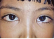
Bilateral eyelids drooping Disease

Blepharophimosis Syndrome (Bilateral eyelid drooping disease)

 Autosomal dominant Condition
(Can be transmitted from both mother or father, usually express in every generation)


Features:

Syndrome consists of bilateral:

– Narrowing of vertical and horizontal palperbral apertures

– Telecanthus

– Inverse epicanthus folds

– Lateral ectropion and moderate to severe ptosis

 May be asymmetrical and without epicanthic folds


 Treatment: 

Plastic reconstruction of lids, along with bilateral brow suspension for ptosis.

Distichiasis

Distichiasis

 Hereditary and congenital condition

 Extra posterior row of cilia, occasionally present in all four lids

 Partial

Complete 


 They occupy the position of meibomian gland orifices

 Eyelashes may irritate cause to corneal epithelial defects

 May be also seen in Stevens Johnson’s syndrome—acquired distichiasis


 Treatment:

By cryotherapy or excision with grafting.

Congenital Lid Conditions: Ophthalmology Spot Diagnosis Series

Coloboma of the Eyelid

 A notch or defect of the lid margin

 Unilateral 

 or bilateral ; upper or lower

 Upper lid coloboma: At the junction of middle and inner thirds

 May be associated with Goldenhar syndrome

 Lower lid coloboma: At middle and outer thirds junction


 Associated with Treacher Collins syndrome

 May be acquired in traumatic cases


 Treatment: 

Urgent plastic repair at a very early age to prevent exposure keratitis and corneal ulcer.

Tuesday, May 10, 2016

Vertigo: what are the causes!

Vertigo

Most important causes of vertigo

DisorderNotes
Viral labyrinthitisRecent viral infection
Sudden onset
Nausea and vomiting
Hearing may be affected
Vestibular neuritisRecent viral infection
Recurrent vertigo attacks lasting hours or days
No hearing loss
Benign paroxysmal positional vertigoGradual onset
Triggered by change in head position
Each episode lasts 10-20 seconds
Meniere's diseaseAssociated with hearing loss, tinnitus and sensation of fullness or pressure in one or both ears
Vertebrobasilar ischaemiaElderly patient
Dizziness on extension of neck
Acoustic neuromaHearing loss, vertigo, tinnitus
Absent corneal reflex is important sign
Associated with neurofibromatosis type 2

Sunday, January 31, 2016

Abdominal Aortic Aneurysm

Abdominal Aortic Aneurysm


Basics
Description
  • Focal dilation of the aortic wall with an increase in diameter by at least 50% (>3 cm)
  • 95% are infrarenal.
  • Gradual expansion or rupture causes symptoms.
  • Rupture can occur into the intraperitoneal or retroperitoneal spaces.
  • Intraperitoneal rupture is usually immediately fatal.
  • Average growth rate of 0.2 to 0.5 cm per year
  • 5-year risk of rupture:
    • Aneurysms <4.0 cm: 2%
    • Aneurysms 4.0-5.0 cm: 5%
    • Aneurysms 5.0-6.0 cm: 25%
    • Aneurysms 6.0-7.0 cm: 35%
  • 40-50% die before they reach the hospital.
  • 50% of patients who reach the hospital alive survive.
  • 5-year survival after repair is 67%.
Geriatric Considerations
  • Risk increases with advanced age.
  • Present in 4-8% of all patients older than 65 years
  • Peak incidence:
    • Men: 5.9% at the age of 80 years
    • Women: 4.5% at the age of 90 years
Etiology
  • Risk factors:
    • Male gender
    • Age >65 years old
    • Family history
    • Cigarette smoking
    • Atherosclerosis
    • Hypertension
    • Diabetes mellitus
    • Connective tissue disorders:
      • Ehlers-Danlos syndrome
      • Marfan syndrome
  • Uncommon causes:
    • Blunt abdominal trauma
    • Infections of the aorta
Related Posts Plugin for WordPress, Blogger...